When the Protein Data Bank Has No Answer: Using AlphaFold2 to Model a Shape-Shifting Virus
Most process models assume the molecule you are separating holds still. For minute virus of mice, that assumption quietly breaks - right in the pH range where anion exchange steps operate.
The consequence is a modeling problem with no obvious solution: the structure you need does not exist in any database. Recent work from Rentschler's Process Science team with KIT solved it by generating the structure instead of looking it up.
A surrogate that is almost, but not quite, the real thing
Virus clearance studies rarely use infectious minute virus of mice (MVM) if they can avoid it. The common stand-in is the mock virus particle (MVP) - a non-infectious, virus-like particle that behaves similarly and is far easier to handle.
The two are not identical, and the difference is structural. MVM's capsid is built from two proteins, VP1 and VP2, in roughly a 1:5 ratio. MVP consists of VP2 only.
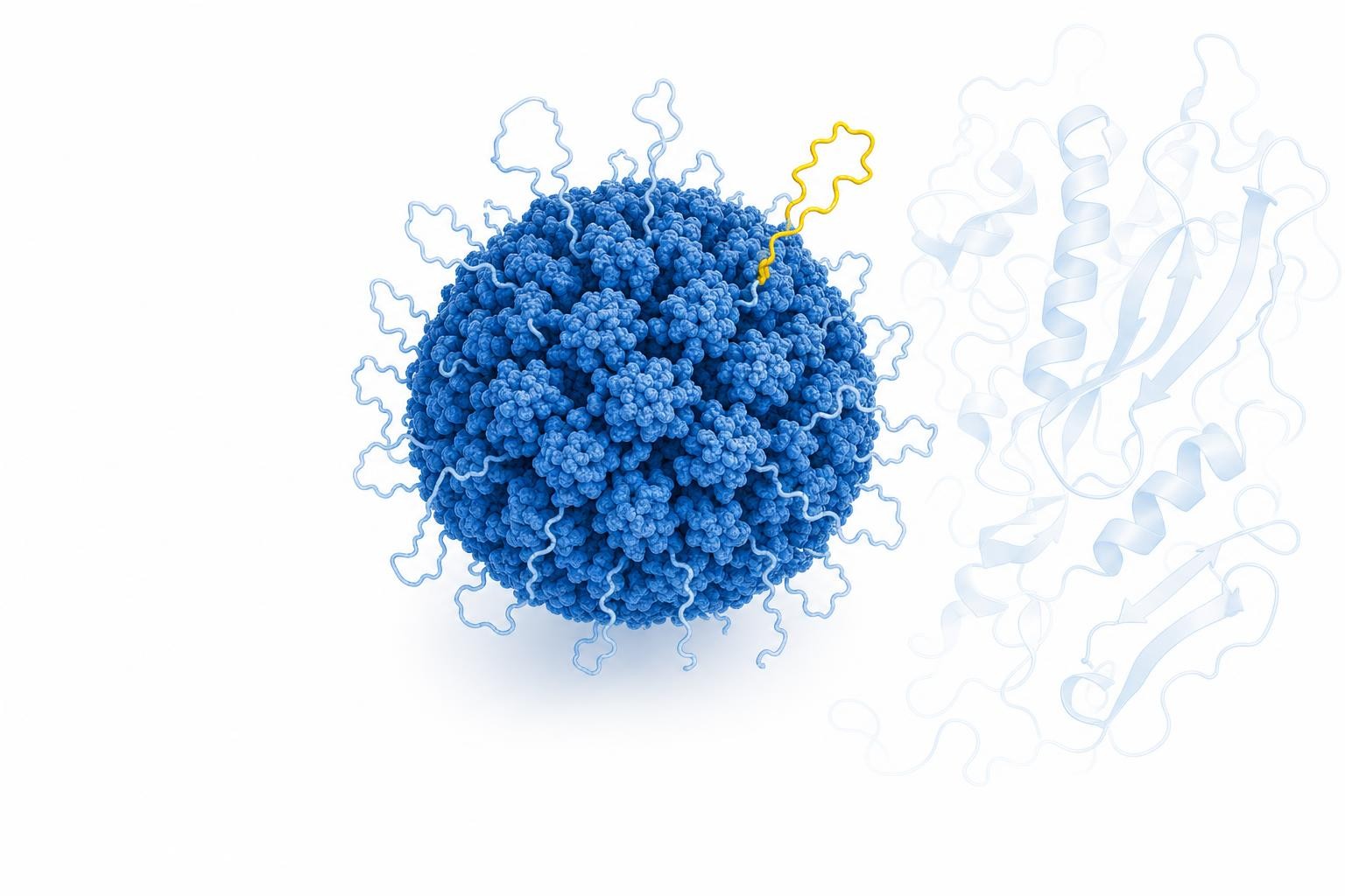
That missing VP1 carries something consequential: an N-terminal phospholipase A2 (PLA2) domain. At neutral to basic pH it sits tucked inside the capsid. Below roughly pH 6, it is externalised - flipped out onto the particle's surface.
For a chromatographer this is not a curiosity. Anion exchange virus removal frequently runs pH gradients that cross that threshold. So partway through the gradient, the particle's surface chemistry changes - and surface chemistry is precisely what determines charge, and therefore retention. It is a plausible explanation for the small but persistent differences in charge and clearance between MVM and MVP reported in the literature.
The structure that isn't there
The workflow underpinning this modeling approach derives a virus's pH-dependent charge from its capsid structure: identify surface-exposed residues, compute a titration curve, correct for the resin phase, fit a polynomial.
That works cleanly for MVP, whose structure is deposited in the Protein Data Bank. For MVM in its post-lysosomal conformation - capsid intact, PLA2 domains externalised - there is no deposited structure. The likely reason is technical: the linker connecting the PLA2 domain to the capsid body is disordered, and disordered regions are notoriously difficult to resolve experimentally.
So the team built one. The full-length VP1 capsid protein was predicted from its amino acid sequence using AlphaFold2, and the resulting top-ranked structure was integrated into the existing capsid at a 1:5 VP1-to-VP2 ratio, with the PLA2 domain placed in its externalised position.
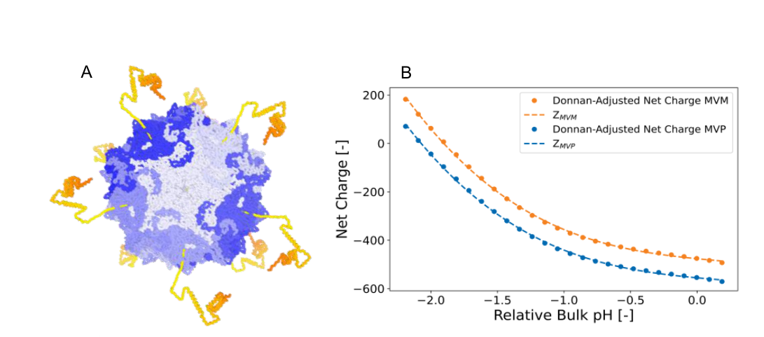
Left: the lysosomal MVM capsid augmented with AlphaFold2-derived externalised PLA2 domains (orange). Right: Donnan-adjusted net charge for MVM compared with MVP.
From there the workflow proceeded exactly as before - SASA to find exposed residues, titration curve across pH, Donnan correction for the pH difference between bulk and resin phase, and a third-order polynomial for charge.
The predicted consequence was visible in the result: the MVM charge curve sits shifted relative to MVP, which is what you would expect when additional charged residues appear on the surface. Encouragingly, that shift was consistent with what earlier virus spiking data had already hinted at.
Zero additional calibration runs
The most economical part of the study is what did not have to be done.
The colloidal particle adsorption model needs two fitted parameters beyond charge: a boundary layer thickness and a kinetic coefficient. For MVM, neither was fitted. Both were transferred directly from the MVP model - identical values - on the reasoning that two particles this structurally similar should share adsorption behaviour once their charge difference is accounted for.
So the MVM model required no new calibration experiments at all. Its charge came from a predicted structure; its remaining parameters came from its surrogate.
That is a meaningful claim in a field where every virus experiment is expensive, and it deserves the scrutiny it invites - which is why the validation against independent MVM spiking data matters. That comparison, including where it agrees and where it doesn't, is covered in the next article in this series.
What this suggests more broadly
The specific result is about one parvovirus on one resin. The transferable idea is larger.
Structure prediction has become good enough that a missing conformation is no longer necessarily a dead end for process modeling. Where a relevant structural state cannot be crystallised - disordered linkers, transient conformations, pH-dependent rearrangements - it may now be possible to generate a defensible working model and carry it into a quantitative framework.
It also reframes the surrogate question. Rather than treating MVP and MVM as interchangeable and absorbing the discrepancy as noise, this approach makes the structural difference explicit and models its consequence. That could improve the comparability of MVP-based spiking studies - one of the more useful outcomes for anyone relying on surrogates in a virus safety strategy.
Based on "Structure based Mechanistic Chromatography Modeling of Mock Virus Particle and Minute Virus of Mice removal using Multi Modal Anion Exchange Chromatography" by Lukas Döring, Thomas Holder, Johannes Winderl, Jonas Kowert and Matthias W. Kron (Process Science, Rentschler Biopharma SE) with Jürgen Hubbuch (Institute of Engineering in Life Sciences, KIT).
Working on a virus clearance strategy? Talk to our Business Development team.